Feline Keratitis and Cats and Review and Fhv-1
Groundwork
Eosinophilic keratitis (EK) is a chronic and progressive keratopathy described in cats and other species, characterized by proliferative white to pink granular corneal plaques and corneal neovascularization (one–5). The verbal pathogenesis of EK is unknown, although an association with feline herpesvirus type one (FHV-1) has been proposed in cats (half-dozen), and the cellular contour is typically consistent with either a blazon I (IgE-mediated) or type Four (T-lymphocyte-mediated) hypersensitivity reaction (7).
Diagnosis of EK can be confirmed with corneal cytology—revealing a predominantly eosinophilic inflammation—while handling of EK is aimed at controlling the immune-mediated disease with the apply of topical and/or systemic allowed-modulatory drugs. Several challenges complicate the clinical direction of EK in cats, including the balance betwixt local immunosuppression and FHV-1 reactivation (viii), hazard of topical corticosteroids in the presence of corneal ulceration (reported in 66.vii% of EK cats) (3), poor therapeutic compliance long-term (2), and relatively high rate of recurrences (upwards to 66%) (1).
The purpose of the present case report is to describe the successful management of severe bilateral EK in a cat, detailing a unique protocol that enhanced therapeutic compliance, improved clinical signs in a rapid fashion, and provided long-term resolution without maintenance immunomodulation (clinical remission at 1 twelvemonth follow upwardly).
Example Presentation
Written informed consent was obtained from the owner for the publication of this case report. A ii-year-quondam male castrated domestic shorthair (vii kg) presented for evaluation of keratitis of approximately 10 months duration. The cat was otherwise good for you with no known systemic diseases, and he was up-to-date on vaccines and flea and tick prevention. An opacity was first noted by the owner in the lateral aspect of the cornea in the left eye (Os), followed weeks afterwards by similar changes in the right eye (OD). The lesions slowly extended over the entire corneal surface in both optics (OU) over several months, resulting in visual impairment, blepharospasm, ocular discharge, and redness. According to the owner, the visual impairment affected the true cat'southward general demeanor (i.due east., hiding beliefs, hands startled) and started disrupting the homo-animal bail in the household. Two months prior to presentation, the cat was prescribed topical 0.1% dexamethasone (Maxidex, Novartis, Basel, Switzerland) by the referring veterinary with a recommended administration frequency of three times daily OU. However, therapy was only given twice daily by the owner due to the patient'due south ocular discomfort and poor compliance, and no improvement was noted at the completion of the 3-calendar week course.
On presentation to a board-certified veterinary ophthalmologist (LS), physical exam and vital parameters were within normal limits. The true cat appeared hesitant in the examination room (difficulty navigating, unable to track cotton balls) and the menace response was absent OU. Dazzle and palpebral reflexes were intact although pupillary lite reflexes were not able to be assessed due to the severity of the corneal opacification OU. Balmy blepharospasm and seromucoid to mucopurulent discharge were present OU. The conjunctiva had diffuse moderate hyperemia and mild chemosis OU. Dense fibrovascular ingrowth affected 100% of the inductive cornea OU, in addition to white raised plaques present on the corneal surface OU (masked past the 3rd eyelid OD) (Figures 1A,B). Ocular diagnostic testing showed that intraocular pressures (TonoVet, Jorgensen Laboratories) were normal (23 mmHg OD, 24 mmHg OS), Schirmer tear examination-i (Merck Fauna Wellness) results were normal (16 mm/min OD, nineteen mm/min Bone) (nine, 10), and multifocal punctate areas of fluorescein uptake were noted OU. Cytological evaluation of the white corneal plaques revealed a mixed, predominately eosinophilic inflammation with few mast cells and lymphocytes (Effigy ii). Clinical diagnoses were severe EK with punctate corneal ulcerations and moderate conjunctivitis OU. Handling was initiated with triamcinolone acetonide (Vetalog, Boehringer Ingelheim Vetmedica, St. Joseph, MO) administered subcutaneously at 0.two mg/kg, too as 0.three% ciprofloxacin ophthalmic solution (Ciloxan, Alcon Laboratories, Fort Worth, TX; 1 drop OU q12h) and two topical compounded medications started v days after upon receipt: 0.5% tacrolimus aqueous solution (Stokes pharmacy, Mount Laurel, NJ; 1 drib OU q6h) and 0.v% cidofovir aqueous solution (Stokes Pharmacy, Mount Laurel, NJ; 1 drop OU q12h).
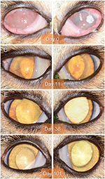
Effigy one. Clinical images of the correct eye (A,C,E,G) and left middle (B,D,F,H) of a two-year-old domestic shorthair cat diagnosed with astringent bilateral eosinophilic keratitis, managed with combined immunosuppressive therapy (subcutaneous triamcinolone acetonide and topical 0.v% tacrolimus), and examined on Day 0 (A,B), Mean solar day 11 (C,D), Day 38 (E,F), and Day 101 (1000,H).
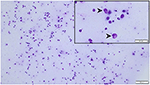
Figure ii. Cytological specimen of the left corneal surface (Wright Giemsa stain) highlighting a mixed, predominantly eosinophilic inflammation (blackness arrowheads) with few mast cells and lymphocytes. Bar = 40 μm (top right insert) or 100 μm (background paradigm).
On Day 1 (i.due east., 24 h following triamcinolone injection), telephonic update with the possessor revealed that the cat'southward visual behavior and ocular comfort were profoundly improved, as well equally the tolerance to handling for topical drug administration. On Day xi, recheck examination showed marked improvement in vision (navigation in exam room, intact menace response OU) and ocular comfort level (no blepharospasm), with notable reduction in ocular belch and conjunctivitis severity OU (mild residual hyperemia). Keratitis was greatly improved with resolution of corneal plaques and epithelial defects OU (no fluorescein uptake), besides equally marked reduction in the fibrovascular ingrowth (mild residue corneal vascularization) assuasive for clear visualization of the within of the eye OU (Figures 1C,D). A subcutaneous injection of triamcinolone acetonide (0.2 mg/kg) was repeated, 0.iii% ciprofloxacin was discontinued, 0.5% cidofovir was continued q12h for ii weeks, and 0.5% tacrolimus was slowly tapered (q8h for 3 weeks then q12h until recheck).
Recheck evaluation on Day 38 revealed resolution of conjunctivitis and ocular belch, and nigh resolution of the keratitis OU (Figures 1E,F). Simply thin superficial vessels were present in the cornea OU, well-nigh of which were hypoperfused ("ghost" vessels). Intraocular pressures (twenty mmHg OD, 25 mmHg OS) and Schirmer tear test-1 results (xx mm/min OD, 25 mm/min OS) were normal. At home handling involved 0.v% tacrolimus at q12h for one week, then q24h for 4 weeks, then q48h with instructions to discontinue administration 1 week before the following recheck.
The true cat was re-examined on Day 101, off any medication for 5 days. The recheck visit confirmed resolution of ocular signs OU with only few hypoperfused superficial corneal vessels noticed on retro-illumination (Figures 1G,H). Throughout all visits, no systemic adverse effects (eastward.chiliad., flare up of herpetic disease such every bit sneezing or nasal discharge) or ocular irritation from compounded tacrolimus were reported by the owner. Further, phone and e-mail communications with the owner confirmed long-term clinical remission (i.e., no recurrence of ocular surface disease) despite discontinuation of all therapies. The latest follow-upward at fourth dimension of manuscript writing was Day 374 (Figures 3, iv).
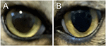
Figure 3. Clinical images of the right eye (A) and left eye (B) of a cat with historical eosinophilic keratitis in clinical remission. Images were taken by the owner using a smartphone on Day 374 following initial diagnosis, that is, 278 days after discontinuation of all therapies.
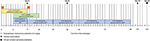
Figure 4. Timeline of the cat's clinical course and treatments.
Give-and-take
The present case report describes the successful management of astringent EK in both eyes of a cat, highlighting two important features: (i) Use of triamcinolone acetonide as the principal method of immunomodulation (rather than conventional topical immunotherapy) to better therapeutic compliance; and (ii) Clinical remission with no recurrence of keratitis despite discontinuation of immunomodulation after simply 3.2 months of therapy.
Triamcinolone acetonide is an intermediate acting glucocorticoid with negligible mineralocorticoid effects, considered approximately 7 times every bit strong as methylprednisone (11). Parenteral administration of triamcinolone provides corticosteroid furnishings for 7–xv days, and a second dose can exist administered post-obit this time if signs persist (12). Hither, triamcinolone acetonide was injected subcutaneously at two visits separated past 11 days, providing a rapid and marked improvement in clinical signs and ocular condolement. Our findings are consistent with a written report of EK in horses, where systemic corticosteroid use was associated with a significantly shorter fourth dimension for resolution of clinical signs (4). Our work is also complementary to the recent study by Lucyshyn et al., in which subcutaneous administration of triamcinolone was accounted prophylactic and as efficacious every bit conventional topical immunomodulation in cats with eosinophilic keratoconjunctivitis (13). All the same, two master differences exist between the two reports: (i) First, clinical remission (noted in our patient) was not described in whatsoever cat reported by Lucyshyn et al., a finding that could be partly explained by the higher intensity of topical therapy in this case study (i.e., high concentration and frequency of tacrolimus); (ii) Second, triamcinolone was administered at time of diagnosis in our feline patient, whereas triamcinolone was preceded by a course of antiviral therapy with famciclovir or 0.5% cidofovir (median 17 days, range 0–63 days) in Lucyshyn'southward written report. The tentative goal of initiating antiviral therapy prior to corticotherapy was to reduce the likelihood of herpetic flare-up and corneal ulceration, even so the authors acknowledged that simultaneous administration of triamcinolone and antiviral drug may be preferred (equally described herein) to reduce fourth dimension to illness resolution, cost, and number of recheck visits.
There are several notable advantages to the unique protocol described in our study. Outset, triamcinolone acetonide provided rapid and pronounced comeback in corneal inflammation, allowing for corneal epithelial defects to heal without the risks associated with topical immunomodulation (i.e., potentiation of infection, delayed re-epithelialization). Second, medication compliance from the patient (and consequently possessor) was profoundly enhanced by reducing ocular discomfort before initiation of topical medications at high frequency; indeed, administration of multiple eyedrops 2–3 times daily (or more) can exist particularly challenging in a cat that is painful and hiding from the possessor. Compliance is particularly relevant for EK as most cases of recurrences were reportedly associated with poor mediation adherence (2). In the present case, the owner struggled medicating the cat prior to the starting time visit, then reported excellent compliance equally early as Day 1 given the substantial improvement in the true cat's ocular condolement and general demeanor. Third, corticosteroid levels on the ocular surface are likely optimized with triamcinolone acetonide equally compared to topical corticosteroid use; the sometime provides a controlled-release formulation that achieves sustained concentrations over an extended menstruation (zero-order kinetics), while the latter provides high concentrations at delivery but rapid turn down due to efficient drug removal via the nasolacrimal drainage appliance (first-order kinetics) (14, 15). Systemic corticosteroid administration achieved quantifiable tear film concentrations in dogs (sixteen), and the same is probable true in cats. Further enquiry is needed to characterize triamcinolone pharmacokinetics in cats, assessing salubrious subjects but also cats with conjunctivitis given the likelihood for college tear film concentrations in eyes with compromised blood-tear barrier (17, 18).
Similar to other reports of cats receiving triamcinolone acetonide for EK or other conditions (13, nineteen), our patient did not experience notable adverse effects from systemic corticotherapy. Nevertheless, this observation should be verified in hereafter prospective studies that include systemic workup and diagnostic testing. Potential agin effects of systemic corticotherapy in cats include (but not limited to) recrudescence of herpetic disease (xx), plasma volume expansion (21) that could promote congestive heart failure in predisposed cats (22), and rarely, iatrogenic hyperadrenocorticism (23).
Maintenance immunotherapy was achieved with topical 0.5% tacrolimus aqueous solution, following an initial frequency of four times daily that was slowly tapered off over 3 months. Of note, the prescribed concentration (0.v%) was much higher than the typical dose of tacrolimus reported in previous ophthalmic studies (0.02%) (24), with the aim to provide an aggressive immunosuppressive therapy from the onset of medical direction (discussed below). Farther, tacrolimus was preferred over previously described 1.5% cyclosporine (2) for two reasons: (i) Tacrolimus is more potent than cyclosporine for reducing corneal neovascularization in patients with severe keratitis, equally reported in dogs with keratoconjunctivitis sicca (24); and (ii) aqueous-based compounded tacrolimus is subjectively ameliorate tolerated than oil-based compounded cyclosporine in cats (authors' personal experience), thus reducing the adventure of stopping therapy due to local adverse furnishings such as marginal blepharitis (2).
Monotherapy is more often than not effective in managing EK in cats, equally described for topical ane.five% cyclosporine (2) or 0.five% megestrol acetate (5), although this arroyo is express by incomplete success rate (11.4 and 12% non-responsive, respectively), relatively irksome clinical improvement (>ii–iii weeks), disease recurrence (22.6 and 33%, respectively), and the need to maintain cats on immunomodulation long-term to lifelong (2, 5). In contrast, the use of combined immunosuppressive drugs in the present case (systemic triamcinolone and topical tacrolimus) achieved a rapid improvement of EK (<11 days) that resulted in clinical remission following discontinuation of all medications by 3.ii months. There is mounting evidence that early on and aggressive immunomodulation improves long-term clinical outcomes and disease prognosis in humans with diverse immune-mediated diseases (e.g., rheumatoid arthritis, Crohn'due south affliction). For instance, human patients with Crohn'due south disease were significantly more likely to achieve clinical remission when receiving combination immunosuppressive therapy (60%) rather than conventional therapy with incremental use of immunomodulation drugs (35.9%) (25). The same may be true in cats with EK given the allowed-mediated pathogenesis of this ocular condition (ii, 5), although the findings of the present case written report should be verified in future prospective controlled studies assessing a larger population of cats.
The main limitation of the study was the lack of diagnostic testing for FHV-1. FHV-ane is detected in over 76% of corneal scrapings from cats with EK (6), and its presence can complicate medical management due to potential herpetic flare upward with immunosuppressive drug. The patient had historical upper respiratory infections during kittenhood, thus 0.five% cidofovir was initiated empirically to reduce the risk for potential FHV-1 re-activation despite the lack of confirmatory testing. Cidofovir was well-tolerated and discontinued 2 weeks after re-epithelialization of corneal ulcers.
Concluding Remarks
The example report highlights the use of combination immunosuppressive therapy to achieve rapid resolution of clinical signs and greater adventure for total recovery (clinical remission). Subcutaneous assistants of triamcinolone acetonide improved ocular condolement and therapeutic compliance—a key component for long-term success—and resulted in excellent disease control in a rapid and effective manner.
Data Availability Argument
The raw data supporting the conclusions of this article will be made available by the authors, without undue reservation.
Ideals Statement
Upstanding review and blessing was not required for the animal written report because the study describes the clinical direction of a patient for a routine ocular condition. Written informed consent was obtained from the owner for the participation of their beast in this study.
Author Contributions
LS examined the patient and conceived the medical direction used in the study. AR and LS wrote the manuscript. All authors contributed to the commodity and canonical the submitted version.
Conflict of Interest
The authors declare that the inquiry was conducted in the absence of whatever commercial or fiscal relationships that could be construed equally a potential conflict of interest.
Acknowledgments
The authors are thankful to the UMN ophthalmology service (Dr. Marina Leis, Stacy Bokelheide, and Melissa Boyd) for their assistance with the patient'southward care.
References
one. Morgan VR. Feline eosinophilic keratitis: a retrospective study of 54 cases:(1989-1994). Vet Comp Ophthalmol. (1996) 6:131–four.
Google Scholar
two. Spiess AK, Sapienza JS, Mayordomo A. Treatment of proliferative feline eosinophilic keratitis with topical 1.5% cyclosporine: 35 cases. Vet Ophthalmol. (2009) 12:132–seven. doi: 10.1111/j.1463-5224.2008.00679.x
PubMed Abstract | CrossRef Full Text | Google Scholar
4. Lassaline-Utter Yard, Miller C, Wotman KL. Eosinophilic keratitis in 46 optics of 27 horses in the Mid-Atlantic The states (2008-2012). Vet Ophthalmol. (2014). 17:311–20. doi: 10.1111/vop.12076
PubMed Abstract | CrossRef Full Text | Google Scholar
five. Stiles J, Coster M. Apply of an ophthalmic formulation of megestrol acetate for the treatment of eosinophilic keratitis in cats. Vet Ophthalmol. (2016) 19(Suppl 1):86–90. doi: 10.1111/vop.12371
PubMed Abstract | CrossRef Full Text | Google Scholar
six. Nasisse MP, Glover TL, Moore CP, Weigler BJ. Detection of feline herpesvirus i DNA in corneas of cats with eosinophilic keratitis or corneal sequestration. Am J Vet Res. (1998) 59:856–8.
PubMed Abstract | Google Scholar
7. Prasse KW. Cytology and histopathology of feline eosinophlic keratitis. Vet Comp Ophthalmol. (1996) six:74–81.
Google Scholar
viii. Nasisse MP. Anti-inflammatory therapy in herpesvirus keratoconjunctivitis. Prog Vet Comp Ophthalmol. (1991) 1:63–5.
PubMed Abstract
ix. Sebbag L, Kass PH, Maggs DJ. Reference values, intertest correlations, and test-retest repeatability of selected tear film tests in healthy cats. J Am Vet Med Assoc. (2015) 246:426–35. doi: ten.2460/javma.246.4.426
PubMed Abstract | CrossRef Total Text | Google Scholar
ten. Sebbag 50, Uhl LK, Schneider B, Hayes B, Olds J, Mochel JP. Investigation of Schirmer tear exam-one for measurement of tear product in cats in diverse environmental settings and with different test durations. J Am Vet Med Assoc. (2020) 256:681–6. doi: ten.2460/javma.256.half-dozen.681
PubMed Abstract | CrossRef Full Text | Google Scholar
eleven. Ganz EC, Griffin CE, Keys DA, Flatgard TA. Evaluation of methylprednisolone and triamcinolone for the induction and maintenance treatment of pruritus in allergic cats: a double-blinded, randomized, prospective study. Vet Dermatol. (2012) 23:387-e72. doi: 10.1111/j.1365-3164.2012.01058.x
PubMed Abstruse | CrossRef Full Text | Google Scholar
12. Ingelheim B. Vetalog Parenteral (Package Insert). St Joseph, MO: Boehringer Ingelheim Vetmedica Inc. (2014).
13. Lucyshyn DR, Expert KL, Knickelbein KE, Chang MW, Strøm AR, Hollingsworth SR, et al. Subcutaneous administration of triamcinolone as part of the management of feline eosinophilic keratoconjunctivitis. J Feline Med Surg. (2020) 2020:1098612X20968660. doi: 10.1177/1098612X20968660
PubMed Abstract | CrossRef Full Text | Google Scholar
14. Sebbag L, Allbaugh RA, Wehrman RF, Uhl LK, Ben-Shlomo Thousand, Chen T, et al. Fluorophotometric assessment of tear book and turnover rate in healthy dogs and cats. J Ocul Pharmacol Ther. (2019) 35:497–502. doi: 10.1089/jop.2019.0038
PubMed Abstract | CrossRef Full Text | Google Scholar
15. Sebbag L, Kirner NS, Allbaugh RA, Reis A, Mochel JP. Kinetics of fluorescein in tear film after middle driblet instillation in beagle dogs: does size really matter? Front Vet Sci. (2019) 6:457. doi: 10.3389/fvets.2019.00457
PubMed Abstract | CrossRef Total Text | Google Scholar
16. Sebbag L, Yan Y, Smith JS, Allbaugh RA, Wulf LW, Mochel JP. Tear fluid pharmacokinetics following oral prednisone administration in dogs with and without conjunctivitis. J Ocul Pharmacol Ther. (2019) 35:341–nine. doi: 10.1089/jop.2019.0020
PubMed Abstract | CrossRef Full Text | Google Scholar
17. Sebbag 50, Allbaugh RA, Weaver A, Seo YJ, Mochel JP. Histamine-induced conjunctivitis and breakdown of claret-tear bulwark in dogs: a model for ocular pharmacology and therapeutics. Front Pharmacol. (2019) x:752. doi: x.3389/fphar.2019.00752
PubMed Abstract | CrossRef Full Text | Google Scholar
18. Sebbag L, Mochel JP. An middle on the domestic dog every bit the scientist's best friend for translational enquiry in ophthalmology: focus on the ocular surface. Med Res Rev. (2020) 40:2566–04. doi: 10.1002/med.21716
PubMed Abstract | CrossRef Full Text | Google Scholar
xix. Momota Y, Yasuda J, Arai Northward, Yamamoto M, Yoshimura H, Ikezawa M, et al. Contribution of oral triamcinolone to treating proliferative and necrotising otitis externa in a 14-year-erstwhile Persian true cat. JFMS Open up Rep. (2017) iii:2055116917691175. doi: 10.1177/2055116917691175
PubMed Abstract | CrossRef Full Text | Google Scholar
xx. Gaskell RM, Povey RC. Re-excretion of feline viral rhinotracheitis virus following corticosteroid treatment. Vet Rec. (1973) 93:204–5. doi: 10.1136/vr.93.seven.204
PubMed Abstract | CrossRef Full Text
21. Block CL, Oyama MA. Echocardiographic and biomarker bear witness of plasma volume expansion afterwards short-term steroids administered orally in cats. J Vet Intern Med. (2020) 34:29–34. doi: 10.1111/jvim.15678
PubMed Abstract | CrossRef Full Text | Google Scholar
22. Smith SA, Tobias AH, Fine DM, Jacob KA, Ployngam T. Corticosteroid-associated congestive heart failure in 12 cats. Intern J Appl Res Vet Med. (2004) two:159–70.
Google Scholar
23. Lien YH, Huang HP, Chang PH. Iatrogenic hyperadrenocorticism in 12 cats. J Am Anim Hosp Assoc. (2006) 42:414–23. doi: 10.5326/0420414
CrossRef Full Text | Google Scholar
24. Radziejewski Chiliad, Balicki I. Comparative clinical evaluation of tacrolimus and cyclosporine eye drops for the treatment of canine keratoconjunctivitis sicca. Acta Vet Hung. (2016) 64:313–29. doi: 10.1556/004.2016.030
PubMed Abstract | CrossRef Full Text | Google Scholar
25. D'Haens G, Baert F, van Assche G, Caenepeel P, Vergauwe P, Tuynman H, et al. Early combined immunosuppression or conventional management in patients with newly diagnosed Crohn's disease: an open randomised trial. Lancet. (2008) 371:660–vii. doi: 10.1016/S0140-6736(08)60304-nine
PubMed Abstract | CrossRef Full Text | Google Scholar
Source: https://www.frontiersin.org/articles/10.3389/fvets.2021.580396/full
0 Response to "Feline Keratitis and Cats and Review and Fhv-1"
Post a Comment